gamma-H2AX (Ser 139) Antibody
- Catalog Number : A05004PC
- Number :
-
Size:
Qty : - Price : Request Inquiry
-


General Information
| Reactivity | Human |
|---|---|
| Application | WB, IF, IHC, ELISA, ChIP |
| Host | Rat |
| Clonality | Polyclonal |
| Conjugate | Non-conjugated |
| Purity | Antigen Affinity Purified |
| Alias | Histone H2AX (H2a/x) (Histone H2A.X), H2AFX, H2AX |
Figure :
|
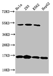
Western Blot Positive WB detected in: Hela whole cell lysate, 293 whole cell lysate, K562 whole cell lysate, HepG2 whole cell lysate All lanes: gamma-H2AX (Ser 139) Antibody at 1.64μg/ml Secondary Goat polyclonal to rabbit IgG at 1/50000 dilution
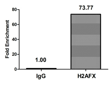
Chromatin Immunoprecipitation Hela (4*106) were treated with Micrococcal Nuclease, sonicated, and immunoprecipitated with 5µg anti-gamma-H2AX (Ser 139) or a control normal rabbit IgG. The resulting ChIP DNA was quantified using real-time PCR with primers against the β-Globin promoter.
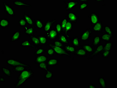
Immunofluorescence staining of Hela cells with gamma-H2AX (Ser 139) Antibody at 1:2.5, counter-stained with DAPI. The cells were fixed in 4% formaldehyde, permeabilized using 0.2% Triton X-100 and blocked in 10% normal Goat Serum. The cells were then incubated with the antibody overnight at 4°C. The secondary antibody was Alexa Fluor 488-congugated AffiniPure Goat Anti-Rabbit IgG(H+L).
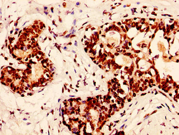
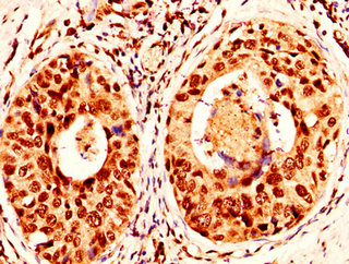
IHC image of gamma-H2AX (Ser 139) Antibody diluted at 1:50 and staining in paraffin-embedded human breast cancer performed on a Leica BondTM system. After dewaxing and hydration, antigen retrieval was mediated by high pressure in a citrate buffer (pH 6.0). Section was blocked with 10% normal goat serum 30min at RT. Then primary antibody (1% BSA) was incubated at 4°C overnight. The primary is detected by a biotinylated secondary antibody and visualized using an HRP conjugated SP system.
IHC image of gamma-H2AX (Ser 139) Antibody diluted at 1:50 and staining in paraffin-embedded human cervical cancer performed on a Leica BondTM system. After dewaxing and hydration, antigen retrieval was mediated by high pressure in a citrate buffer (pH 6.0). Section was blocked with 10% normal goat serum 30min at RT. Then primary antibody (1% BSA) was incubated at 4°C overnight. The primary is detected by a biotinylated secondary antibody and visualized using an HRP conjugated SP system. |


.png)