
JAK1 (phospho Tyr1034) Polyclonal Antibody
- Catalog Number : A130154
- Number : A130154
-
Size:
Qty : - Price : Request Inquiry
-


General Information
| Reactivity | Human, Mouse, Rat | |||||||||
|---|---|---|---|---|---|---|---|---|---|---|
| Application | WB, IF, IHC, ELISA, IP | |||||||||
| Host | Rabbit | |||||||||
| Clonality | Polyclonal | |||||||||
| Conjugate | Non-conjugated | |||||||||
| Isotype | IgG | |||||||||
| Immunogen | The antiserum was produced against synthesized peptide derived from human JAK1 around the phosphorylation site of Tyr1034. AA range:988-1037 | |||||||||
| Molecular Weight | 132kD (Observed) | |||||||||
| Storage buffer | Liquid in PBS containing 50% glycerol, 0.5% BSA and 0.02% sodium azide. | |||||||||
| Storage instruction | -15°C to -25°C/1 year(Do not lower than -25°C) | |||||||||
| Research topic | >>EGFR tyrosine kinase inhibitor resistance>>PI3K-Akt signaling pathway>>Necroptosis | |||||||||
| Alias | JAK1
JAK1A JAK1B Tyrosine-protein kinase JAK1 Janus kinase 1 JAK-1 |
|||||||||
| Recommended Dilution Ratio | IF 1:50-200; WB 1:200-1:1000; IHC 1:100-1:300; ELISA 1:10000; Not yet tested in other applications | |||||||||
| Specificity | Phospho-JAK1 (Y1034) Polyclonal Antibody detects endogenous levels of JAK1 protein only when phosphorylated at Y1034.The name of modified sites may be influenced by many factors, such as species (the modified site was not originally found in human samples) and the change of protein sequence (the previous protein sequence is incomplete, and the protein sequence may be prolonged with the development of protein sequencing technology). When naming, we will use the "numbers" in historical reference to keep the sites consistent with the reports. The antibody binds to the following modification sequence (lowercase letters are modification sites):KEyYT | |||||||||
| Purification | The antibody was affinity-purified from rabbit antiserum by affinity-chromatography using epitope-specific immunogen. | |||||||||
| Gene Name | JAK1 | |||||||||
| Protein Name | Tyrosine-protein kinase JAK1 | |||||||||
| Database Link |
| |||||||||
| Background | This gene encodes a membrane protein that is a member of a class of protein-tyrosine kinases (PTK) characterized by the presence of a second phosphotransferase-related domain immediately N-terminal to the PTK domain. The encoded kinase phosphorylates STAT proteins (signal transducers and activators of transcription) and plays a key role in interferon-alpha/beta and interferon-gamma signal transduction. Alternative splicing results in multiple transcript variants. [provided by RefSeq, Mar 2016]. | |||||||||
| Function | Catalytic activity:ATP + a [protein]-L-tyrosine = ADP + a [protein]-L-tyrosine phosphate.,Domain:Possesses two phosphotransferase domains. The second one probably contains the catalytic domain (By similarity), while the presence of slight differences suggest a different role for domain 1.,Domain:The FERM domain mediates interaction with JAKMIP1.,Function:Tyrosine kinase of the non-receptor type, involved in the IFN-alpha/beta/gamma signal pathway. Kinase partner for the interleukin (IL)-2 receptor.,sequence Caution:Translation N-terminally extended.,similarity:Belongs to the protein kinase superfamily. Tyr protein kinase family. JAK subfamily.,similarity:Contains 1 FERM domain.,similarity:Contains 1 protein kinase domain.,similarity:Contains 1 SH2 domain.,subcellular location:Wholly intracellular, possibly membrane associated.,subunit:Interacts with IL31RA, JAKMIP1 and SHB.,tissue specificity:Expressed at higher levels in primary colon tumors than in normal colon tissue. The expression level in metastatic colon tumors is comparable to the expression level in normal colon tissue. | |||||||||
| Cellular Localization | Endomembrane system; Peripheral membrane protein. Wholly intracellular, possibly membrane associated. | |||||||||
| Tissue Expression | Expressed at higher levels in primary colon tumors than in normal colon tissue. The expression level in metastatic colon tumors is comparable to the expression level in normal colon tissue. | |||||||||
| Validation Data |
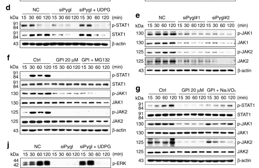
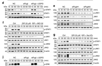
Ma, J., Wei, K., Liu, J. et al. Glycogen metabolism regulates macrophage-mediated acute inflammatory responses. Nat Commun 11, 1769 (2020). | |||||||||
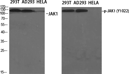
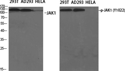
Western Blot analysis of various cells using Phospho-JAK1 (Y1022) Polyclonal Antibody diluted at 1:500 | ||||||||||
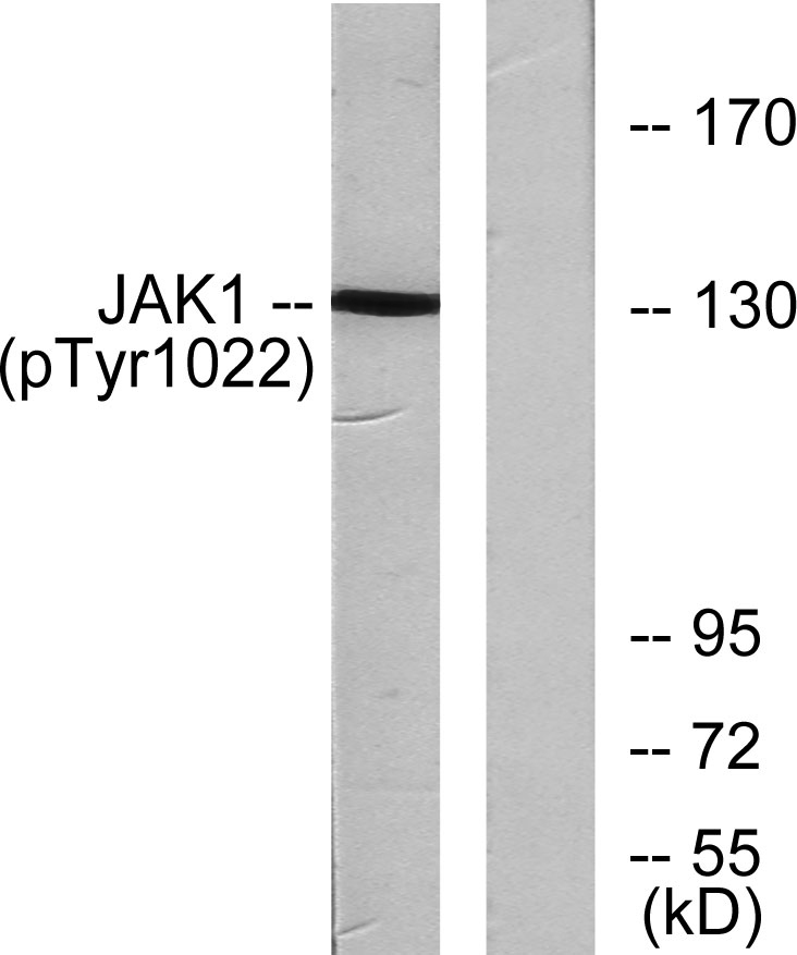
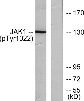
Western blot analysis of lysates from A549 cells , using JAK1 (Phospho-Tyr1022) Antibody. The lane on the right is blocked with the phospho peptide. | ||||||||||
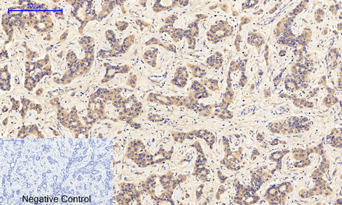
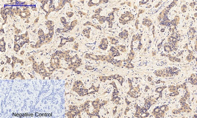
Immunohistochemical analysis of paraffin-embedded Human-liver-cancer tissue. 1,JAK1 (phospho Tyr1022) Polyclonal Antibody was diluted at 1:200(4°C,overnight). 2, Sodium citrate pH 6.0 was used for antibody retrieval(>98°C,20min). 3,Secondary antibody was diluted at 1:200(room tempeRature, 30min). Negative control was used by secondary antibody only. | ||||||||||
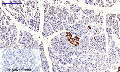
Immunohistochemical analysis of paraffin-embedded Human-stomach-cancer tissue. 1,JAK1 (phospho Tyr1022) Polyclonal Antibody was diluted at 1:200(4°C,overnight). 2, Sodium citrate pH 6.0 was used for antibody retrieval(>98°C,20min). 3,Secondary antibody was diluted at 1:200(room tempeRature, 30min). Negative control was used by secondary antibody only. | ||||||||||
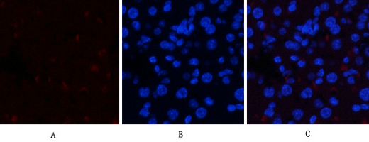
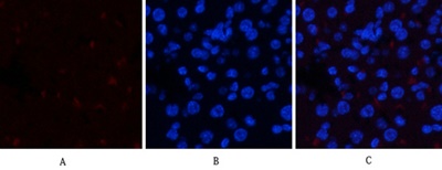
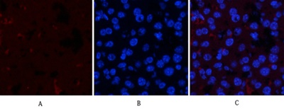
Immunofluorescence analysis of mouse-liver tissue. 1,JAK1 (phospho Tyr1022) Polyclonal Antibody(red) was diluted at 1:200(4°C,overnight). 2, Cy3 labled Secondary antibody was diluted at 1:300(room temperature, 50min).3, Picture B: DAPI(blue) 10min. Picture A:Target. Picture B: DAPI. Picture C: merge of A+B | ||||||||||
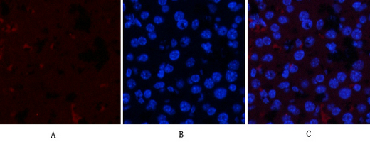
Immunofluorescence analysis of mouse-liver tissue. 1,JAK1 (phospho Tyr1022) Polyclonal Antibody(red) was diluted at 1:200(4°C,overnight). 2, Cy3 labled Secondary antibody was diluted at 1:300(room temperature, 50min).3, Picture B: DAPI(blue) 10min. Picture A:Target. Picture B: DAPI. Picture C: merge of A+B |


.png)