
JAK3 Rabbit pAb
- Catalog Number : A12430
- Number : A12430
-
Size:
Qty : - Price : Request Inquiry
-


General Information
| Reactivity | Human, Mouse, Rat | ||||||||||||
|---|---|---|---|---|---|---|---|---|---|---|---|---|---|
| Application | WB, IF, IHC, ELISA | ||||||||||||
| Host | Rabbit | ||||||||||||
| Clonality | Polyclonal | ||||||||||||
| Conjugate | Non-conjugated | ||||||||||||
| Isotype | IgG | ||||||||||||
| Immunogen | The antiserum was produced against synthesized peptide derived from human JAK3. AA range:751-800 | ||||||||||||
| Molecular Weight | 125kD (Observed) | ||||||||||||
| Storage buffer | Liquid in PBS containing 50% glycerol, 0.5% BSA and 0.02% sodium azide. | ||||||||||||
| Storage instruction | -15°C to -25°C/1 year(Do not lower than -25°C) | ||||||||||||
| Research topic | >>Chemokine signaling pathway>>PI3K-Akt signaling pathway>>Necroptosis | ||||||||||||
| Alias | JAK3
Tyrosine-protein kinase JAK3 Janus kinase 3 JAK-3 Leukocyte janus kinase L-JAK |
||||||||||||
| Recommended Dilution Ratio | WB 1:500-1:2000; IHC 1:100-1:300; IF 1:200-1:1000; ELISA 1:20000; Not yet tested in other applications. | ||||||||||||
| Specificity | JAK3 Polyclonal Antibody detects endogenous levels of JAK3 protein. | ||||||||||||
| Purification | The antibody was affinity-purified from rabbit antiserum by affinity-chromatography using epitope-specific immunogen. | ||||||||||||
| Gene Name | JAK3 | ||||||||||||
| Protein Name | Tyrosine-protein kinase JAK3 | ||||||||||||
| Database Link |
| ||||||||||||
| Background | The protein encoded by this gene is a member of the Janus kinase (JAK) family of tyrosine kinases involved in cytokine receptor-mediated intracellular signal transduction. It is predominantly expressed in immune cells and transduces a signal in response to its activation via tyrosine phosphorylation by interleukin receptors. Mutations in this gene are associated with autosomal SCID (severe combined immunodeficiency disease). [provided by RefSeq, Jul 2008]. | ||||||||||||
| Function | Catalytic activity:ATP + a [protein]-L-tyrosine = ADP + a [protein]-L-tyrosine phosphate.,Disease:Defects in JAK3 are a cause of severe combined immunodeficiency autosomal recessive T-cell-negative/B-cell-positive/NK-cell-negative (T(-)B(+)NK(-)SCID) [MIM:600802]. SCID refers to a genetically and clinically heterogeneous group of rare congenital disorders characterized by impairment of both humoral and cell-mediated immunity, leukopenia, and low or absent antibody levels. Patients with SCID present in infancy with recurrent, persistent infections by opportunistic organisms. The common characteristic of all types of SCID is absence of T-cell-mediated cellular immunity due to a defect in T-cell development.,Domain:Possesses two phosphotransferase domains. The second one probably contains the catalytic domain (By similarity), while the presence of slight differences suggest a different role for domain 1.,Function:Tyrosine kinase of the non-receptor type, involved in the interleukin-2 and interleukin-4 signaling pathway. Phosphorylates STAT6, IRS1, IRS2 and PI3K.,online information:JAK3 mutation db,PTM:Tyrosine phosphorylated in response to IL-2 and IL-4.,similarity:Belongs to the protein kinase superfamily. Tyr protein kinase family. JAK subfamily.,similarity:Contains 1 FERM domain.,similarity:Contains 1 protein kinase domain.,similarity:Contains 1 SH2 domain.,subcellular location:Wholly intracellular, possibly membrane associated.,subunit:Interacts with STAM2 and MYO18A (By similarity). Interacts with SHB.,tissue specificity:In NK cells and an NK-like cell line but not in resting T-cells or in other tissues. The S-form is more commonly seen in hematopoietic lines, whereas the B- and M-forms are detected in cells both of hematopoietic and epithelial origins. | ||||||||||||
| Cellular Localization | Endomembrane system ; Peripheral membrane protein. Cytoplasm. | ||||||||||||
| Tissue Expression | In NK cells and an NK-like cell line but not in resting T-cells or in other tissues. The S-form is more commonly seen in hematopoietic lines, whereas the B-form is detected in cells both of hematopoietic and epithelial origins. | ||||||||||||
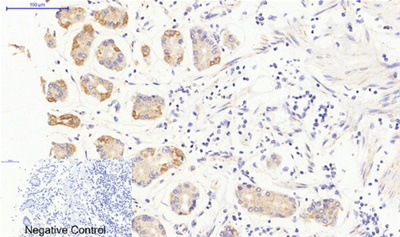
| Validation Data |
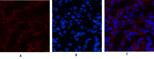
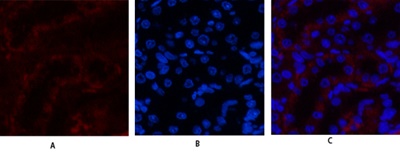
Immunofluorescence analysis of rat-kidney tissue. 1,JAK3 Polyclonal Antibody(red) was diluted at 1:200(4° overnight). 2, Cy3 labled Secondary antibody was diluted at 1:300(room temperature, 50min).3, Picture B: DAPI(blue) 10min. Picture A:Target. Picture B: DAPI. Picture C: merge of A+B | ||||||||||||
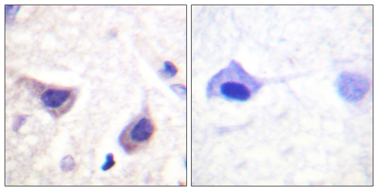
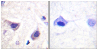
Immunohistochemistry analysis of paraffin-embedded human brain tissue, using JAK3 Antibody. The picture on the right is blocked with the synthesized peptide. | |||||||||||||
Immunofluorescence analysis of rat-kidney tissue. 1,JAK3 Polyclonal Antibody(red) was diluted at 1:200(4° overnight). 2, Cy3 labled Secondary antibody was diluted at 1:300(room temperature, 50min).3, Picture B: DAPI(blue) 10min. Picture A:Target. Picture B: DAPI. Picture C: merge of A+B |


.png)