
STAT3 (Phospho Tyr705) (PT0742R) PT® Rabbit mAb
- Catalog Number : AM8551
- Number : AM8551
-
Size:
Qty : - Price : Request Inquiry
-


General Information
| Reactivity | Human, Mouse, Rat | |||||||||
|---|---|---|---|---|---|---|---|---|---|---|
| Application | WB, IF, IHC, ELISA, IP | |||||||||
| Host | Rabbit | |||||||||
| Clonality | Monoclonal | |||||||||
| Conjugate | Non-conjugated | |||||||||
| Isotype | IgG,Kappa | |||||||||
| Molecular Weight | 88kD (Calculated) | 88kD (Observed) | |||||||||
| Storage buffer | PBS, 50% glycerol, 0.05% Proclin 300, 0.05%BSA | |||||||||
| Storage instruction | -15°C to -25°C/1 year(Do not lower than -25°C) | |||||||||
| Research topic | >>EGFR tyrosine kinase inhibitor resistance>>Chemokine signaling pathway>>HIF-1 signaling pathway | |||||||||
| Alias | STAT3 | |||||||||
| Recommended Dilution Ratio | IHC 1:200-1:1000; WB 1:2000-1:10000; IF 1:200-1:1000; ELISA 1:5000-1:20000; IP 1:50-1:200 | |||||||||
| Specificity | Phospho-Stat3 (Y705) protein detects endogenous levels of STAT3 | |||||||||
| Purification | Protein A | |||||||||
| Gene Name | STAT3 | |||||||||
| Protein Name | STAT3 | |||||||||
| Database Link |
| |||||||||
| Background | The protein encoded by this gene is a member of the STAT protein family. In response to cytokines and growth factors, STAT family members are phosphorylated by the receptor associated kinases, and then form homo- or heterodimers that translocate to the cell nucleus where they act as transcription activators. This protein is activated through phosphorylation in response to various cytokines and growth factors including IFNs, EGF, IL5, IL6, HGF, LIF and BMP2. This protein mediates the expression of a variety of genes in response to cell stimuli, and thus plays a key role in many cellular processes such as cell growth and apoptosis. The small GTPase Rac1 has been shown to bind and regulate the activity of this protein. PIAS3 protein is a specific inhibitor of this protein. Mutations in this gene are associated with infantile-onset multisystem autoimmune disease and hyper. | |||||||||
| Function | Disease:Defects in STAT3 are the cause of hyperimmunoglobulin E recurrent infection syndrome autosomal dominant (AD-HIES) [MIM:147060]; also known as hyper-IgE syndrome or Job syndrome. AD-HIES is a rare disorder of immunity and connective tissue characterized by immunodeficiency, chronic eczema, recurrent Staphylococcal infections, increased serum IgE, eosinophilia, distinctive coarse facial appearance, abnormal dentition, hyperextensibility of the joints, and bone fractures.,Function:Transcription factor that binds to the interleukin-6 (IL-6)-responsive elements identified in the promoters of various acute-phase protein genes. Activated by IL31 through IL31RA.,miscellaneous:Involved in the gp130-mediated signaling pathway.,online information:STAT3 entry,online information:STAT3 mutation db,PTM:Tyrosine phosphorylated in response to IL-6, IL-11, CNTF, LIF, CSF-1, EGF, PDGF, IFN-alpha and OSM. Phosphorylated on serine upon DNA damage, probably by ATM or ATR. Serine phosphorylation is important for the formation of stable DNA-binding STAT3 homodimers and maximal transcriptional activity.,similarity:Belongs to the transcription factor STAT family.,similarity:Contains 1 SH2 domain.,subcellular location:Shuttles between the nucleus and the cytoplasm. Constitutive nuclear presence is independent of tyrosine phosphorylation.,subunit:Forms a homodimer or a heterodimer with a related family member (at least STAT1). Interacts with NCOA1, PELP1, SOCS7 and STATIP1. Interacts with HCV core protein. Interacts with IL23R in presence of IL23. Interacts with IL31RA. Interacts with SIPAR. Interacts (via SH2 domain) with NLK (By similarity). Interacts with KPNA4 and KPNA5; KPNA4 may be the primary mediator of nuclear import (By similarity). Interacts with TMF1.,tissue specificity:Heart, brain, placenta, lung, liver, skeletal muscle, kidney and pancreas. | |||||||||
| Cellular Localization | Cytoplasm. Nucleus. Shuttles between the nucleus and the cytoplasm. Translocated into the nucleus upon tyrosine phosphorylation and dimerization, in response to signaling by activated FGFR1, FGFR2, FGFR3 or FGFR4. Constitutive nuclear presence is independent of tyrosine phosphorylation. Predominantly present in the cytoplasm without stimuli. Upon leukemia inhibitory factor (LIF) stimulation, accumulates in the nucleus. The complex composed of BART and ARL2 plays an important role in the nuclear translocation and retention of STAT3. Identified in a complex with LYN and PAG1. | |||||||||
| Tissue Expression | Heart, brain, placenta, lung, liver, skeletal muscle, kidney and pancreas. Expressed in naive CD4(+) T cells as well as T-helper Th17, Th1 and Th2 cells (PubMed:31899195). | |||||||||
| Validation Data |
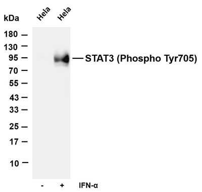
Various whole cell lysates were separated by 4-20% SDS-PAGE, and the membrane was blotted with anti-STAT3 (Phospho Tyr705) (PT0742R) antibody. The HRP-conjugated Goat anti-Rabbit IgG(H + L) antibody was used to detect the antibody. Lane 1: Hela Lane 2: Hela was starved of serum overnight and then treated with INF-α(100 ng/mL) for 5 minutes Predicted band size: 88kDa Observed band size: 88kDa | |||||||||
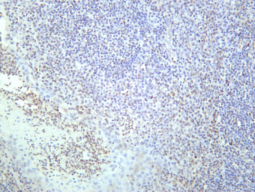
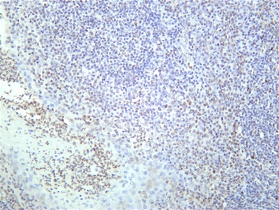
Human tonsil was stained with anti-STAT3 (Phospho Tyr705) rabbit antibody | ||||||||||
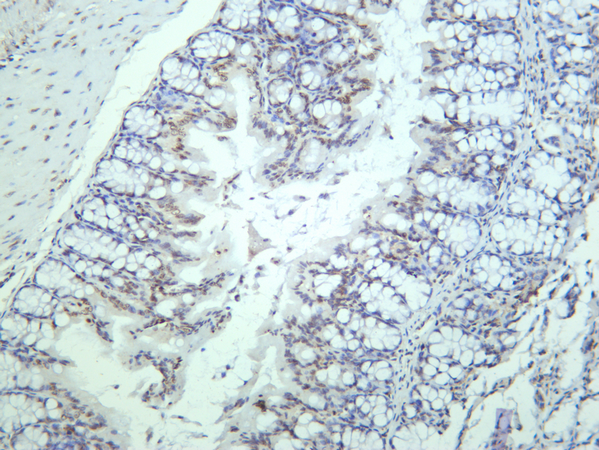
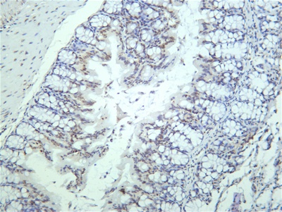
Mouse colon was stained with anti-STAT3 (Phospho Tyr705) rabbit antibody | ||||||||||
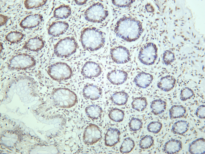
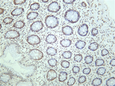
Human colon was stained with anti-STAT3 (Phospho Tyr705) rabbit antibody |


.png)