


Stat3 (Phospho Ser727) Rabbit pAb
- Catalog Number : AP0250
- Number : AP0250
-
Size:
Qty : - Price : Request 詢價
- Stock : Request
-


General Information
| Reactivity | Human, Mouse, Rat, Monkey | ||||||||||||
|---|---|---|---|---|---|---|---|---|---|---|---|---|---|
| Application | WB, IF, IHC, ELISA, IP | ||||||||||||
| Host | Rabbit | ||||||||||||
| Clonality | Polyclonal | ||||||||||||
| Conjugate | Non-conjugated | ||||||||||||
| Isotype | IgG | ||||||||||||
| Immunogen | The antiserum was produced against synthesized peptide derived from human STAT3 around the phosphorylation site of Ser727. AA range:694-743 | ||||||||||||
| Molecular Weight | 85kD (Observed) | ||||||||||||
| Storage buffer | Liquid in PBS containing 50% glycerol, 0.5% BSA and 0.02% sodium azide. | ||||||||||||
| Storage instruction | -15°C to -25°C/1 year(Do not lower than -25°C) | ||||||||||||
| Research topic | >>EGFR tyrosine kinase inhibitor resistance>>Chemokine signaling pathway>>HIF-1 signaling pathway | ||||||||||||
| Alias | STAT3
APRF Signal transducer and activator of transcription 3 Acute-phase response factor |
||||||||||||
| Recommended Dilution Ratio | IF 1:50-200; WB 1:500-1:2000; IHC 1:100-1:300; IP 2-5 ug/mg lysate; ELISA 1:10000; Not yet tested in other applications | ||||||||||||
| Specificity | Phospho-Stat3 (S727) Polyclonal Antibody detects endogenous levels of Stat3 protein only when phosphorylated at S727.The name of modified sites may be influenced by many factors, such as species (the modified site was not originally found in human samples) and the change of protein sequence (the previous protein sequence is incomplete, and the protein sequence may be prolonged with the development of protein sequencing technology). When naming, we will use the "numbers" in historical reference to keep the sites consistent with the reports. The antibody binds to the following modification sequence (lowercase letters are modification sites):PMsPR | ||||||||||||
| Purification | The antibody was affinity-purified from rabbit antiserum by affinity-chromatography using epitope-specific immunogen. | ||||||||||||
| Gene Name | STAT3 | ||||||||||||
| Protein Name | Signal transducer and activator of transcription 3 | ||||||||||||
| Database Link |
| ||||||||||||
| Background | The protein encoded by this gene is a member of the STAT protein family. In response to cytokines and growth factors, STAT family members are phosphorylated by the receptor associated kinases, and then form homo- or heterodimers that translocate to the cell nucleus where they act as transcription activators. This protein is activated through phosphorylation in response to various cytokines and growth factors including IFNs, EGF, IL5, IL6, HGF, LIF and BMP2. This protein mediates the expression of a variety of genes in response to cell stimuli, and thus plays a key role in many cellular processes such as cell growth and apoptosis. The small GTPase Rac1 has been shown to bind and regulate the activity of this protein. PIAS3 protein is a specific inhibitor of this protein. Mutations in this gene are associated with infantile-onset multisystem autoimmune disease and hyper | ||||||||||||
| Function | Disease:Defects in STAT3 are the cause of hyperimmunoglobulin E recurrent infection syndrome autosomal dominant (AD-HIES) [MIM:147060]; also known as hyper-IgE syndrome or Job syndrome. AD-HIES is a rare disorder of immunity and connective tissue characterized by immunodeficiency, chronic eczema, recurrent Staphylococcal infections, increased serum IgE, eosinophilia, distinctive coarse facial appearance, abnormal dentition, hyperextensibility of the joints, and bone fractures.,Function:Transcription factor that binds to the interleukin-6 (IL-6)-responsive elements identified in the promoters of various acute-phase protein genes. Activated by IL31 through IL31RA.,miscellaneous:Involved in the gp130-mediated signaling pathway.,online information:STAT3 entry,online information:STAT3 mutation db,PTM:Tyrosine phosphorylated in response to IL-6, IL-11, CNTF, LIF, CSF-1, EGF, PDGF, IFN-alpha and OSM. Phosphorylated on serine upon DNA damage, probably by ATM or ATR. Serine phosphorylation is important for the formation of stable DNA-binding STAT3 homodimers and maximal transcriptional activity.,similarity:Belongs to the transcription factor STAT family.,similarity:Contains 1 SH2 domain.,subcellular location:Shuttles between the nucleus and the cytoplasm. Constitutive nuclear presence is independent of tyrosine phosphorylation.,subunit:Forms a homodimer or a heterodimer with a related family member (at least STAT1). Interacts with NCOA1, PELP1, SOCS7 and STATIP1. Interacts with HCV core protein. Interacts with IL23R in presence of IL23. Interacts with IL31RA. Interacts with SIPAR. Interacts (via SH2 domain) with NLK (By similarity). Interacts with KPNA4 and KPNA5; KPNA4 may be the primary mediator of nuclear import (By similarity). Interacts with TMF1.,tissue specificity:Heart, brain, placenta, lung, liver, skeletal muscle, kidney and pancreas. | ||||||||||||
| Cellular Localization | Cytoplasm . Nucleus . Shuttles between the nucleus and the cytoplasm. Translocated into the nucleus upon tyrosine phosphorylation and dimerization, in response to signaling by activated FGFR1, FGFR2, FGFR3 or FGFR4. Constitutive nuclear presence is independent of tyrosine phosphorylation. Predominantly present in the cytoplasm without stimuli. Upon leukemia inhibitory factor (LIF) stimulation, accumulates in the nucleus. The complex composed of BART and ARL2 plays an important role in the nuclear translocation and retention of STAT3. Identified in a complex with LYN and PAG1. | ||||||||||||
| Tissue Expression | Heart, brain, placenta, lung, liver, skeletal muscle, kidney and pancreas. Expressed in naive CD4(+) T cells as well as T-helper Th17, Th1 and Th2 cells (PubMed:31899195). | ||||||||||||
| Validation Data |
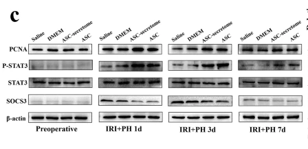
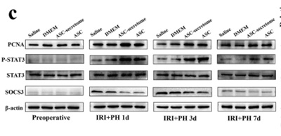
Jiao, Z., Ma, Y., Zhang, Q. et al. The adipose-derived mesenchymal stem cell secretome promotes hepatic regeneration in miniature pigs after liver ischaemia-reperfusion combined with partial resection. Stem Cell Res Ther 12, 218 (2021). | ||||||||||||
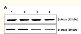
Tang, Qiusha, et al. "Combination of PEI-Mn0. 5Zn0. 5Fe2O4 nanoparticles and pHsp 70-HSV-TK/GCV with magnet-induced heating for treatment of hepatoma." International journal of nanomedicine 10 (2015): 7129. | |||||||||||||
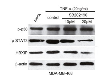
Cai, Xiaoli, et al. "Inflammatory factor TNF-α promotes the growth of breast cancer via the positive feedback loop of TNFR1/NF-κB (and/or p38)/p-STAT3/HBXIP/TNFR1." Oncotarget 8.35 (2017): 58338. | |||||||||||||
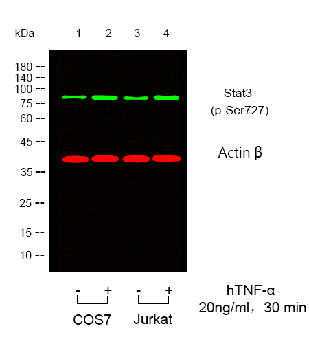
Western blot analysis of lysates from COS7,Jurkat cells, treated or untreated with TNFa, (Green)primary antibody was diluted at 1:1000, 4°over night, secondary antibody was diluted at 1:10000, 37° 1hour. (Red) loading contrl antibody was diluted at 1:5000 as loading control, 4° over night,secondary antibody was diluted at 1:10000, 37° 1hour. | |||||||||||||
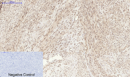
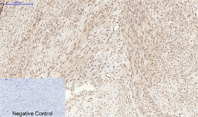
Immunohistochemical analysis of paraffin-embedded Human-uterus tissue. 1,Stat3 (phospho Ser727) Polyclonal Antibody was diluted at 1:200(4°C,overnight). 2, Sodium citrate pH 6.0 was used for antibody retrieval(>98°C,20min). 3,Secondary antibody was diluted at 1:200(room tempeRature, 30min). Negative control was used by secondary antibody only. | |||||||||||||
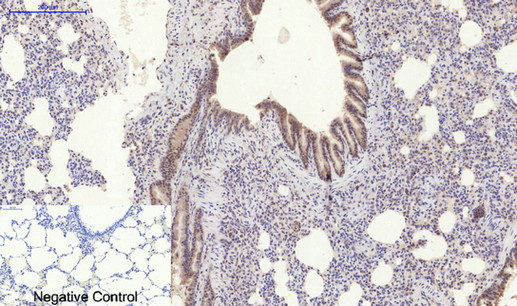
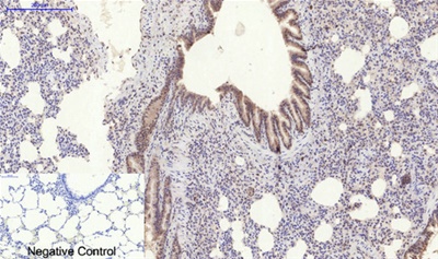
Immunohistochemical analysis of paraffin-embedded Rat-lung tissue. 1,Stat3 (phospho Ser727) Polyclonal Antibody was diluted at 1:200(4°C,overnight). 2, Sodium citrate pH 6.0 was used for antibody retrieval(>98°C,20min). 3,Secondary antibody was diluted at 1:200(room tempeRature, 30min). Negative control was used by secondary antibody only. | |||||||||||||
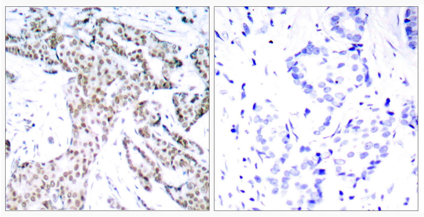
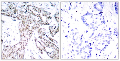
Immunohistochemistry analysis of paraffin-embedded human breast carcinoma, using STAT3 (Phospho-Ser727) Antibody. The picture on the right is blocked with the phospho peptide. | |||||||||||||
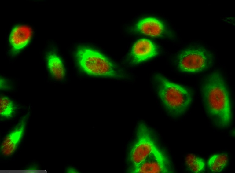
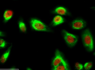
Immunofluorescence analysis of Hela cell. 1,Stat3 (phospho Ser727) Polyclonal Antibody(red) was diluted at 1:200(4° overnight). Caspase 9 Monoclonal Antibody(3-20)(green) was diluted at 1:200(4° overnight). 2, Goat Anti Rabbit Alexa Fluor 594 Catalog:RS3611 was diluted at 1:1000(room temperature, 50min). Goat Anti Mouse Alexa Fluor 488 Catalog:RS3208 was diluted at 1:1000(room temperature, 50min). |


.png)